
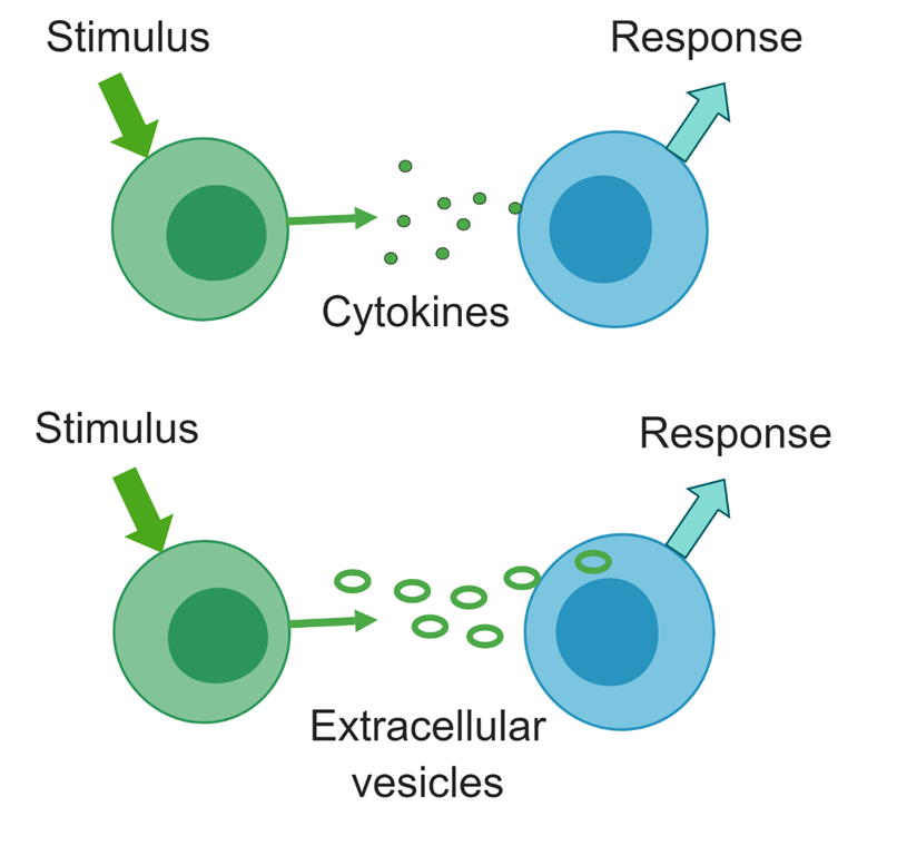
 Cells signal each other through release and uptake of cytokines and extracellular vesicles (EVs). The three types of EVs are exosomes, produced through exocytosis, microvesicles, which bud from the cell membrane, and apoptotic bodies, which are products of dying cells. EVs carry both proteins such as cytokines and microRNAs, which can influence gene expression and activity of recipient cells. We are isolating EVs from ME/CFS patients and healthy controls and examining their protein, metabolite, and RNA cargo.
This project is funded through our NIH ME/CFS Center and more information can be found on the Center website:
https://neuroimmune.cornell.edu/research/vesicles-and-signaling/
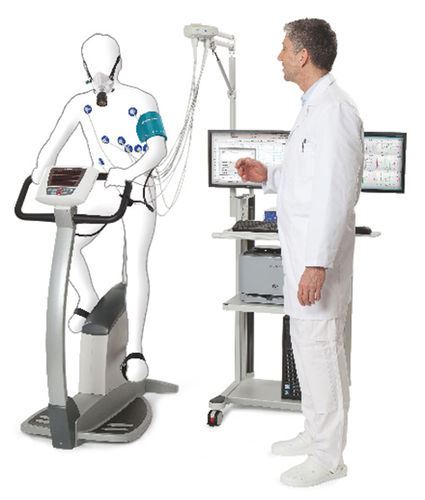
 In collaboration with Dr. Betsy Keller in the School of Health Sciences and Human Performance at Ithaca College, we are examining the physiological and molecular basis of the exercise intolerance characteristics of CFS/ME patients. We are examining the effect of a maximal exercise stress test on gene expression, metabolism, inflammatory markers, and cytokine levels in ME/CFS patients and healthy controls.
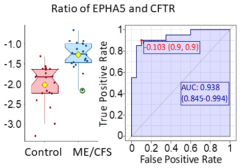
The ratio of the protein EPHA5 and CFTR gives a high level of discrimination between ME/CFS patients and controls, according to ROC analysis. Both of the proteins are involved in regulation of neurotransmitter release in response to varying blood glucose concentrations.
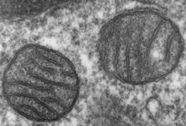
Dysfunction of mitochondria, the energy-generating organelle in human cells, is one hypothesis concerning the severe fatigue experienced by ME/CFS patients. To investigate whether there are genetic differences in the mitochondrial DNA of patients and healthy controls, we have sequenced the mitochondrial genome of a cohort of subjects recruited by the Chronic Fatigue Initiative.
 A publication for this project was published in the Journal of Translational Medicine:
Mitochondrial DNA variants correlate with symptoms in myalgic encephalomyelitis/chronic fatigue syndrome.
by Paul Billing-Ross, Arnaud Germain, Kaixiong Ye, Alon Keinan, Zhenglong Gu, and Maureen R. Hanson
J. Translational Medicine. 2016, 14:19
Click here for a simplified explanation of the JTM publication.
 Many individuals with ME/CFS describe digestive disturbances. These symptoms might result from altered composition of the gut microbiome, as is known in intestinal inflammatory diseases. We have characterized the gut bacterial and eukaryotic microbiome in a cohort of ME/CFS patients vs. healthy controls, using 16S rRNA sequencing and 18S rRNA sequencing, respectively. We have observed a greater bacterial diversity in the ME/CFS patients and have observed differences in the taxonomic composition of the microbiomes of individuals and of the two populations.
Results of the study are available here: